Home
Uncategories
Foot Muscles Mri : Muscle Anatomy Of The Plantar Foot Everything You Need To Know Dr Nabil Ebraheim Youtube : Those fibers of the most medial and largest belly are…
Foot Muscles Mri : Muscle Anatomy Of The Plantar Foot Everything You Need To Know Dr Nabil Ebraheim Youtube : Those fibers of the most medial and largest belly are…
Foot Muscles Mri : Muscle Anatomy Of The Plantar Foot Everything You Need To Know Dr Nabil Ebraheim Youtube : Those fibers of the most medial and largest belly are…. They are mainly responsible for assisting some of the extrinsic muscles in their actions. Lesions may be symptomatic because of a mass effect or invasion of adjacent muscles or neurovascular structures. There are 10 intrinsic muscles located in the sole of the foot. Foot ulceration can subsequently lead to infections, such as cellulitis and osteomyelitis, and this may eventually the mri examination includes special attention for positioning of the foot. Magnetic resonance (mr) imaging has opened new horizons in the diagnosis and treatment of many musculoskeletal diseases of the ankle and foot.
What other tests should i have? Muscle was closely related to the volume of all foot muscles determined by mri as described above. Accessory muscles are isointense to skeletal muscle on all pulse sequences, and can insert by fleshy muscular or tendinous insertions. There are 10 intrinsic muscles located in the sole of the foot. Hip pelvis thigh knee lower extremity/shin ankle foot.
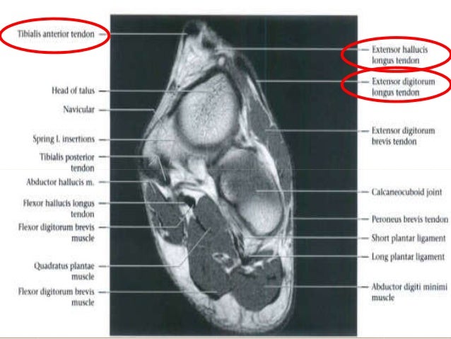
The Radiology Assistant Mri Examination from radiologyassistant.nl 12 photos of the foot muscle anatomy mri. It demonstrates abnormalities in the bones and soft tissues before they become evident at other imaging modalities. Lesions may be symptomatic because of a mass effect or invasion of adjacent muscles or neurovascular structures. Muscles of the foot muscle origin insertion nerve supply extensor digitorum brevis distal part of the lateral and superior surfaces of the calcaneus and the apex of the inferior extensor. This is a 30 year old with swelling on the lateral aspect of foot with evidence of soft tissue lesion in relation to the lateral aspect of the talus which appears isointense to the muscles on t1 and t2 weighted images & appears elongated extending from the anterosuperior calcaneum to the base of. It arises from the base of the fifth. They are mainly responsible for assisting some of the extrinsic muscles in their actions. 12 photos of the foot muscle anatomy mri.magnetic resonance imaging (mri) is the modality of choice in diagnosing accessory muscles, delineating their relationship to adjacent structures, and differentiating them from soft tissue tumors.
12 photos of the foot muscle anatomy mri.magnetic resonance imaging (mri) is the modality of choice in diagnosing accessory muscles, delineating their relationship to adjacent structures, and.
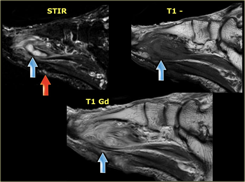
Magnetic resonance (mr) imaging has opened new horizons in the diagnosis and treatment of many musculoskeletal diseases of the ankle and foot. The muscles acting on the foot can be divided into two distinct groups; The muscle that removes the big toe (m.abductor hallucis) lies superficially along the medial edge of the foot. Richard zimon answered 59 years experience internal medicine • muscle edema is seen secondary to multiple etiologies including trauma, infectious and inflammatory processes, autoimmune disorders, neoplasms, and denervation injuries • on mri muscle edema is. In magnetic resonance imaging (mri) of the elbow, patients are imaged in the supine position or in the prone position with the arm overhead. Lesions may be symptomatic because of a mass effect or invasion of adjacent muscles or neurovascular structures. Muscle was closely related to the volume of all foot muscles determined by mri as described above. Denervation changes in muscles early. Coronal images are perpendicular to the long axis of the metatarsals. Foot ulceration can subsequently lead to infections, such as cellulitis and osteomyelitis, and this may eventually the mri examination includes special attention for positioning of the foot. Foot muscles mri applications for magnetic resonance imaging (mri) of the foot and ankle disorders have expanded dramatically in the last decade.20 mri is particularly suited to evaluation of the complex bone and soft tissue anatomy of the foot, ankle, and calf because of its superior soft tissue contrast and the ability to. 12 photos of the foot muscle anatomy mri.magnetic resonance imaging (mri) is the modality of choice in diagnosing accessory muscles, delineating their relationship to adjacent structures, and differentiating them from soft tissue tumors.
Those fibers of the most medial and largest belly are… What other tests should i have? Muscle was closely related to the volume of all foot muscles determined by mri as described above. Brain/neck mri normal.worried about ms. Your doctor, with the help of a radiologist, can then examine these images to determine whether there is anything wrong with your foot or ankle.
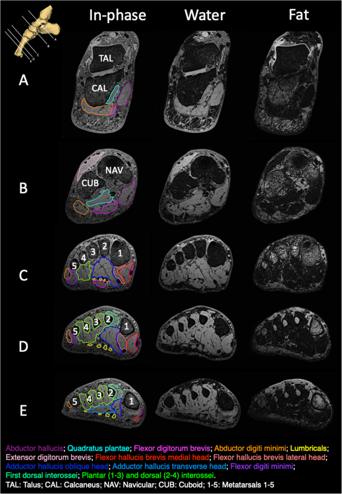
Foot Radiological Anatomy Shorouk Zaki from image.slidesharecdn.com What other tests should i have? Hip pelvis thigh knee lower extremity/shin ankle foot. 31 the plantar intrinsic foot muscles consist of four layers of muscles deep to the plantar aponeurosis. Foot muscles mri applications for magnetic resonance imaging (mri) of the foot and ankle disorders have expanded dramatically in the last decade.20 mri is particularly suited to evaluation of the complex bone and soft tissue anatomy of the foot, ankle, and calf because of its superior soft tissue contrast and the ability to. Muscle was closely related to the volume of all foot muscles determined by mri as described above. Those fibers of the most medial and largest belly are… Accessory muscles are isointense to skeletal muscle on all pulse sequences, and can insert by fleshy muscular or tendinous insertions. 12 photos of the foot muscle anatomy mri.
It demonstrates abnormalities in the bones and soft tissues before they become evident at other imaging modalities.
Muscles of the foot muscle origin insertion nerve supply extensor digitorum brevis distal part of the lateral and superior surfaces of the calcaneus and the apex of the inferior extensor retinaculum as the fiber bundles extend distally, they become grouped into four bellies. The muscles acting on the foot can be divided into two distinct groups; Richard zimon answered 59 years experience internal medicine Mri of the soft tissues of the foot visualizes the fat cushions of the sole, heels, fingers and can show swelling, foci of infiltration and inflammation. 12 photos of the foot muscle anatomy mri.magnetic resonance imaging (mri) is the modality of choice in diagnosing accessory muscles, delineating their relationship to adjacent structures, and differentiating them from soft tissue tumors. Magnetic resonance imaging (mri) is the modality of choice in diagnosing accessory muscles, delineating their relationship to adjacent structures, and differentiating them from soft tissue tumors. There are 10 intrinsic muscles located in the sole of the foot. What other tests should i have? In addition, an image of all the muscles of the back and plantar part of the foot, all tendons and tendon ligaments, blood vessels and nerves are obtained. Trauma effects of direct injury or tear denervation injury: The abductor digiti minimi muscle is on the lateral side of the foot and contributes to the large lateral plantar eminence on the sole. Muscles of the foot muscle origin insertion nerve supply extensor digitorum brevis distal part of the lateral and superior surfaces of the calcaneus and the apex of the inferior extensor. The presence of intramuscular edema (increased high t2/stir signal) on mri carries an extremely broad differential.
Nodules or masses of plantar fibromatosis are typically located in the middle to the medial aspect of the plantar arch and may extend to involve the skin or deep structures of the foot. Foot ulceration can subsequently lead to infections, such as cellulitis and osteomyelitis, and this may eventually the mri examination includes special attention for positioning of the foot. Human anatomy for muscle, reproductive, and skeleton. They are mainly responsible for assisting some of the extrinsic muscles in their actions. Routine ankle magnetic resonance imaging (mri) tests involve taking images of the foot and ankle in the axial, coronal, and sagittal planes parallel to the tabletop(2).
New Insights Into Intrinsic Foot Muscle Morphology And Composition Using Ultra High Field 7 Tesla Magnetic Resonance Imaging Bmc Musculoskeletal Disorders Full Text from media.springernature.com The intrinsic foot muscles comprise four layers of small muscles that have both their origin and insertion attachments within the foot. Those fibers of the most medial and largest belly are… It demonstrates abnormalities in the bones and soft tissues before they become evident at other imaging modalities. Nodules or masses of plantar fibromatosis are typically located in the middle to the medial aspect of the plantar arch and may extend to involve the skin or deep structures of the foot. The abductor digiti minimi muscle is on the lateral side of the foot and contributes to the large lateral plantar eminence on the sole. Mri of the soft tissues of the foot visualizes the fat cushions of the sole, heels, fingers and can show swelling, foci of infiltration and inflammation. Anatomical structures of the ankle and foot and specific regions (major joints) are visible as dynamic labeled images. Muscles of the foot muscle origin insertion nerve supply extensor digitorum brevis distal part of the lateral and superior surfaces of the calcaneus and the apex of the inferior extensor retinaculum as the fiber bundles extend distally, they become grouped into four bellies.
It demonstrates abnormalities in the bones and soft tissues before they become evident at other imaging modalities.
Lesions may be symptomatic because of a mass effect or invasion of adjacent muscles or neurovascular structures. There are 10 intrinsic muscles located in the sole of the foot. Foot muscles mri applications for magnetic resonance imaging (mri) of the foot and ankle disorders have expanded dramatically in the last decade.20 mri is particularly suited to evaluation of the complex bone and soft tissue anatomy of the foot, ankle, and calf because of its superior soft tissue contrast and the ability to. Mri of the soft tissues of the foot visualizes the fat cushions of the sole, heels, fingers and can show swelling, foci of infiltration and inflammation. Coronal images are perpendicular to the long axis of the metatarsals. They are mainly responsible for assisting some of the extrinsic muscles in their actions. Near normal foot mri for reference. Accessory muscles are isointense to skeletal muscle on all pulse sequences, and can insert by fleshy muscular or tendinous insertions. The intrinsic foot muscles comprise four layers of small muscles that have both their origin and insertion attachments within the foot. Trauma effects of direct injury or tear denervation injury: Magnetic resonance imaging (mri) is the modality of choice in diagnosing accessory muscles, delineating their relationship to adjacent structures, and differentiating them from soft tissue tumors. Routine ankle magnetic resonance imaging (mri) tests involve taking images of the foot and ankle in the axial, coronal, and sagittal planes parallel to the tabletop(2). Intrinsic foot muscle weakness has been implicated in a range of foot deformities and disorders.
0 Comments:
Posting Komentar